Tampilkan postingan dengan label Hemoglobin. Tampilkan semua postingan

Jumat, 29 Agustus 2014

Structures relevant to liganded hemoglobins

a. Stereochemistry of the Active Site Before the advent of techniques that enabled the preparation and stabilization of oxyhemoglobin crystals, key information on the probable structure of oxyhemoglobin and thence on the mechanism of cooperativity was extrapolated from structures of methemoglobin derivatives II and from various five- and six-coordinate cobalt-porphyrinato complexes. llO,112,113 The structures of these met derivatives have proved to be similar to that of oxyhemoglobin, at least in the stereochemistry of the metalloporphyrinato species and for the protein tertiary and quaternary structure as well. 
Two, synthetic iron-dioxygen adducts built from the picket-fence porphyrins have been structurally characterized. 172,18? The high, effectively fourfold symmetry of the binding pocket in these systems results in fourfold disorder of the angularly coordinated dioxygen molecule, and precludes the precise and accurate measurements of the Fe-O-O angle and 0-0 separation that are grist to the theoretical mills. * Figure 4.28B illustrates the stereochemistry for one conformer. Subsequently, the structures of several dioxygen adducts of biolog-


ical oxygen carriers have been determined.183.188-191 Although the dioxygen moiety is usually ordered, the precision is tantalizingly just less than that needed to decide whether the apparently more-linear geometry seen for oxyerythrocruorin 183 and oxyhemoglobin 190 is significantly different from that for oxymyoglobin 188 and therefore attributable to the water molecule or imidazole that is hydrogenbonded to the coordinated dioxygen ligand. Nonetheless, several interesting differences emerge.
The axial base in oxymyoglobin and oxyhemoglobin is almost eclipsed; that is, </>1 = 0°. The axial base has moved from a tilted position in deoxyhemoglobin to a symmetric one in oxyhemoglobin. In the absence of steric constraints, the iron atom is essentially in the center of the porphyrin plane for Fe(PF)( 1MeIm)( 02), oxymyoglobin, and oxyhemoglobin. For the 2-methyl analogue, Fe(PF)(2-MeIm)(02), the iron remains significantly out of the plane, as also appears to occur for oxyerythrocruorin.
In the structure of Fe(TPP)(Py)(CO), Figure 4.30, a model for carbonyl hemoglobins, the iron atom is in the plane and the Fe-C == 0 bond is linear and perpendicular, as expected. 118 Not so for carbonyl hemoglobins, where the blob of electron density that is identified with the coordinated carbon monoxide lies substantially off the normal to the porphyrin. We return to this point shortly.
In general, with the exception of the coordinated ligand, the structures of sixcoordinate low-spin hemoglobins, whether FeU or Fe III, are similar. Indeed, the refined structures of oxy- and carbonmonoxyhemoglobin are superimposable within experimental uncertainties, except in the immediate vicinity of the diatomic ligand. Some metrical details are given in Table 4.8.


b. Interactions of Coordinated Ligands with Distal Groups Without exception to date (but see footnote 1 in Reference 168), in structurally characterized oxyhemoglobins, the coordinated dioxygen ligand is hydrogen-bonded to the distal histidine or to a water molecule--even though theoretical calculations show that hydrogen bonding would destabilize M-02 moieties. 192 This universal observation of hydrogen bonding in these biological systems is consistent with notions that electron density accumulates on the dioxygen molecule upon coordination. Given the errors associated with atomic positions (at best, ± 0.20 A) the x-ray crystallographic evidence could be equivocal, since hydrogen atoms on the distal imidazole are not observed. There are at least three lines of evidence that support the existence of a specific O2 ... HN interaction. First, the EPR spectrum of cobalt oxyhemoglobin indicates that the coordinated dioxygen is hydrogen-bonded to something. 177 - 179 Second, and more directly, in the neutron- diffraction structure of oxymyoglobin,189 where hydrogen and especially deuterium nuclides scatter strongly, the imino hydrogen or deuteron was located on the nitrogen atom closest to the coordinated dioxygen, as illustrated in Figure 4.31A. In contrast, in the neutron-diffraction structure of carbonmonoxymyoglobin, the alternative imidazole tautomer was observed (Figure 4.3IB). 125,189 The absence of hydrogen bonding of the distal imidazole residue with the coordinated CO molecule is consistent with other lines of evidence that there is little accumulation of electron density on the carbonyl ligand. Third, but less directly, genetically engineered mutants have been produced in which the distal histidine has been replaced by glycine-sperm whale Mb E7His-....? Gly, and HbA Q'E7His-....?Gly and HbA /3E7His-....?Gly.35b,192 For the myoglobin mutant, the O2 binding rate constant at room temperature increases by an order of magnitude, but the dissociation rate constant increases by two orders of magnitude, leading to a decrease in affinity of more than an order of magnitude, as derived from kon/koff' This leads to an estimate of the free energy associated with hydrogen bonding of
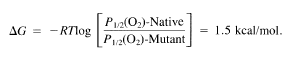


In addition, this mutant myoglobin autooxidizes rapidly compared to the native one. On the other hand, the affinity for CO is greatly increased, leading to a value of M for the mutant of 1300, compared to 16 for the native. Thus the distal histidine stabilizes a coordinated O2 ligand by hydrogen bonding and destabilizes a coordinated CO ligand by steric clash.
A similar discrimination is seen for the Q' chains of the hemoglobin mutant in the binding of the fourth O2 or CO molecule. For the /3 chains little difference is seen relative to the native protein: hydrogen bonding between the distal histidine and the coordinated dioxygen ligand appears to be much weaker in /3 chains, as evidenced by longer N(H) ... 0 separations than those seen in the Q' chains. Comparison of the crystal structures of the native and mutant Q'2(/3E7His-....? GlYh structures reveals negligible changes in the distal environ


 ment, except for that occasioned by the replacement of -CHZ-C3NzH3 (histidine side chain) by -H (glycine side chain). Studies of hemoglobin mutants where the nonpolar distal residue ,BValEll (-CH(CH3h) is replaced by alanine (-CH3), isoleucine (-CH(CH3)CHzCH3), and leucine (-CHzCH(CH3h) reveal that this valine offers steric hindrance to oxygen binding in the T state.

Whereas the angularly coordinated O2 ligand fits comfortably around the distal histidine, a perpendicular and linear CO moiety cannot. Either the distal histidine rotates out of the way, or the CO tilts off axis, or the Fe-C == 0 group bends, or some combination of these occurs. Notwithstanding the absence of bent M-CO moieties in the inorganic literature, reports of strongly bent M-CO groups appear in the biochemical literature. 122-127 The controversy is illustrative of the synergistic interplay of data from models and proteins, and the importance of examining a problem with a miscellany of techniques. The molecular orbital model of ligand-metal interactions presented in Figure 4.22 does not preclude a bent M-C==O moiety on symmetry grounds. Groups related to CO can bend; the normally linearly coordinated SCN - moiety has been observed 194 to become strongly bent under severe steric stress, with an Fe-N-CS of 140°. Unfortunately, the resolution in protein crystal structures is not sufficient to distinguish unequivocally a linear tilted stereochemistry from a bent one or from a combination of tilt and bend. Studies by the XANES technique have been interpreted in terms of a bent Fe-C == 0 moiety (150°)

both in MbCO 127 and in the CO adduct of a chelated heme in micelles, the latter being an especially surprising result. From EXAFS data on a number of carbonyl adducts, two interpretations were offered: linear or moderately bent (150°) FeCO moieties for unhindered model systems, and moderately bent or strongly bent (130°) FeCO moieties for hindered synthetic and biological systems. 195 In the crystal structure of MbCO at 1.5 Aresolution,122 the CO group is disordered, and Fe-C == 0 angles of 120° and 140° were proposed, although the alternative model of tilted, nearly linear Fe-C == 0 stereochemistry could not be eliminated, and is indeed far more likely to account for the off-axis nature of the oxygen position. Vibrational spectroscopy confirms the existence of two major configurations, and indicates a third minor configuration of the Fe-C == 0 moiety in MbCO. 196 An elegant infrared study of the polarization of reattached carbon-monoxide molecules following photolysis of MbCO by linearly polarized light at 10 K gave tilt angles of the CO vector with respect to the heme normal of 15(3t, 28(2)°, and 33(4t for the three conformational substates; 196b the former two values were confirmed in a similar study at room temperature. 196c Note that these studies do not yield the tilt of the Fe-C bond to the heme normal.
 
In three synthetic compounds with severe steric hindrance, the extent of bending and tilting of the Fe-CO moiety is small. In one nonporphyrinic system the Fe-C==O group is bent by 9.4(5t and tilted by 4.20.121a In Fe(PocPF)( l,2-Me2Im)(CO) the Fe-C== 0 angle is 172.5(6t and modest tilting of the Fe-CO group and substantial buckling of the porphyrin ring are apparent. 121b In Fe(C2Cap)((1-MeIm)(CO) the two independent Fe-C==O angles are 172.9(6t and 175.9(6t and modest tilting of the Fe-CO group is again apparent.

From a detailed analysis of the force constants describing the vibrational spectroscopy for the Fe-CO moiety, values of 171 Q for the Fe-CO angle, 9.5° for the tilt, and 11° for porphyrin buckling were calculated for MbCO. l21c These results are particularly important, for in a model complex very closely related to Fe(Poc-PF)(l,2-Me2Im)(CO), just mentioned, an EXAFS study 195 suggested an Fe-C == 0 bond angle of 127(4t; that same study ascribed an Fe-C == 0 bond angle of around 130° to MbCO. The structure of carbonmonoxyhemoglobin, Hb(CO)4' now is interpreted in terms of a nearly linear tilted geometry. 192 Clearly the geometry of attachment of CO to hemoglobins is perturbed by the surroundings of the ligand-binding site and hence the affinity of hemoglobins for CO is also perturbed. Unfortunately, a clear resolution of the geometry of the Fe-CO moiety in MbCO does not exist yet.

Structures relevant to deoxy hemoglobins

The structure of the picket-fence porphyrin compound, Fe(PF)(2-MeIm), is shown in Figure 4.28. 172 Minus the pickets, it is essentially a magnified view of the active site of deoxymyoglobin, shown in Figure 4.29. 181 Some metrical details of these structures, of a very similar unsubstituted tetraphenylporphyrin, I 10 and of several other deoxyhemoglobins I Ie, 182-185 are listed in Table 4.7. In general they are all similar, but important differences exist.
 
In all structures, except deoxyerythrocruorin,183 the iron atom is displaced about 0.4 to 0.5 Afrom the plane of the porphyrin toward the axial base. For deoxyerythrocruorin the displacement is less than half this, perhaps because the water molecule is weakly coordinated to the iron center.


An imidazole group from a histidine residue-the distal histidine E7 in position 7 on helix labeled E-hovers over the binding site for most vertebrate hemoglobins, except for genetically engineered mutants of human hemoglobin (j3E7His -> Gly), pathological mutant hemoglobins, such as hemoglobin Zurich (j3E7His -> Arg), and some others, such as elephant hemoglobin. Long believed to be noncoordinating, this distal histidine may, in fact, coordinate weakly to the Fe center at low temperature. 159 In the a chains of human deoxyhemoglo-


bin, hemoglobin A, a water molecule is found in the binding cavity. 182 For many years the binding cavity has been referred to as the hydrophobic pocketliterally, water-hating. Although many hydrophobic groups, such as valine, leucine, isoleucine, and phenylalanine are positioned over the porphyrin, the immediate environment around the binding site is, in fact, polar, with the distal histidine and associated water molecules, as well as the heme group itself. As will be shown in the next section, the label "hydrophobic pocket" becomes more misleading when the interaction of coordinated ligands with distal groups is examined.
 
The orientation of the axial base, angle 1>1, is similar for Fe(PF)(2-MeIm) and for several vertebrate deoxyhemoglobins. On the other hand, Fe(TPP)(2Melm) and deoxyerythrocruorin have a similar eclipsed axial-base orientation. At least for five-coordinate species, where the iron center is substantially out of the porphyrin plane, orientation of the axial base does not invariably induce structural perturbations, e.g., doming, in the porphyrin skeleton. The conformation of the protein chain is such that the proximal histidine in
deoxyhemoglobin coordinates in a slightly tilted manner,182,186 comparable to the tilt that the sterically active 2-methyl substituent induces in the synthetic systems. 172 Clearly, coordination of the histidine to the heme in a symmetric manner, as would be expected in the absence of the protein constraints, does not produce the conformation of lowest free energy for the whole molecule.

Ligand Affinities in Hemoglobins and Their Models

The O2 affinities in biological carriers span five orders of magnitude, which at room temperature corresponds to a difference in the free energy of oxygen binding


of about 6.0 kcallmol. This wide range of O2 and CO affinities has not yet been paralleled in synthetic systems; the values for O2 affinity do not exceed those for R-state human hemoglobin. A selection of values from model systems is given in Table 4.5. 23 ,31,160-]65 For the flat-open porphyrin system (Figure 4.23) the dioxygen ostensibly binds in an unconstrained manner, but is actually subject to solvent influences. In order to obtain thermodynamic constants on these "unhindered" systems, one must gather data at several low temperatures and then extrapolate to room temperature, or obtain them from kinetic measurements, K = kon/koff, at room temperature.
For the picket-fence porphyrins, dioxygen binds in a protected pocket that is deep enough to accommodate it and to prevent the dimerization that leads to irreversible oxidation, provided that there is a slight excess of base to ensure full saturation of the coordination sites on the unprotected face of the porphyrin. 72 Thus the picket-fence, the capped, and the bis-pocket porphyrins reversibly bind dioxygen at room temperature with little oxidation over many cycles.
This stability facilitated isolation of crystals of a synthetic iron-dioxygen species of the picket-fence porphyrin. The capped porphyrin offers a more highly protected site. The low affinity these latter systems have for dioxygen indicates that the binding cavity is so small that repulsive steric interactions between coordinated dioxygen and the cap are unavoidable. The left-hand side of Figure 4.24 depicts on a logarithmic scale the range of O2 affinities. Each power of 10 corresponds to around 1.2 kcallmol at 25°C.
The right-hand side of Figure 4.24 illustrates the range of affinities for CO binding. For many synthetic systems the CO affinities are orders of magnitude greater than in the biological systems that have an O2 affinity similar to the synthetic; for example, see the entries for the picket-fence porphyrin. Comparison of the left- and right-hand sides of Figure 4.24 reveals that the strongest O2 binder, hemoglobin Ascaris, is one of the weakest CO binders. The O2 affinity of the picket-fence porphyrins is very similar to that of myoglobin, but, as will be detailed shortly, one cannot infer from this that the binding sites are strictly comparable. Indeed, similar affinities have been observed with a non-porphyrin iron complex. 121 ,162 Moreover, if the CO affinity of myoglobin paralleled that of the picket-fence porphyrins, some 20 percent of myoglobin (and hemoglobin) would be in the carbonmonoxy form (in contrast to the approximately 3 percent that occurs naturally), a level that could render reading this section while chewing gum physically taxing.


There is a convenient index to summarize the extent to which CO (or O2) binding is discriminated against for a given iron-porphyrin system. M is defined as the ratio of O2 affinity (as P1/2) to CO affinity for a particular system and experimental conditions:


From Figure 4.24 and from Tables 4.2 and 4.5 the M values calculated may be somewhat arbitrarily divided into three classes: those where M > 2 X 10 4 (good CO binder); those where 2 x 10 2 < M < 2 X 10⁴ ; and those where M < 2 X 10 2 (good O2 binder). An analogous parameter, N, may be defined to summarize the differences in the O2 affinity between an iron-porphyrin system and its cobalt analogue:


For the picket-fence porphyrins and for vertebrate hemoglobins N is in the range 10 to 250, whereas for the flat-open porphyrins and for some hemoglobins that lack a distal histidine (e.g., hemoglobin Glycera and hemoglobin Aplysia) , N is at least an order of magnitude larger, indicating for these latter species that the cobalt analogue binds O2 relatively poorly 167,168 (see Table 4.6).
Note that whereas the O2 binding of the picket-fence porphyrins is similar to that for myoglobin, the kinetics of the process are very different; the synthetic system is more than an order of magnitude faster in kl and k_ 1 (often also referred to as kon and koff)' On the other hand, O2 binding to the pocket porphyrin is similar to that for the biological system. The factors by which ligand affinities are modulated, generally to the benefit of the organism, are subtle and varied, and their elucidation requires the precise structural information that is currently available only from x-ray diffraction experiments. Figure 4.25 shows the structural features of interest that will be elaborated upon in the next subsections.

Kamis, 28 Agustus 2014

The hemoglobin family

Hemoglobins are the most evolutionarily diverse family of dioxygen carriers. They are found in some plants (e.g., leghemoglobin in the nitrogen-fixing nodules of legumes), many invertebrates (including some insect larvae), crustaceans, molluscs (especially bivalves and snails), almost all annelid worms, and in all vertebrates with one possible exception, the Antarctic fish Cyclostomata. With few exceptions the monomeric and oligomeric hemoglobins all share a basically similar building block: a single heme group is embedded in a folded polypeptide with a molecular weight of about 16 kDa (see Figure 4.2), and is anchored to the protein by coordination of the iron center to an imidazole ligand from a histidine residue. Mammalian myoglobin is often taken as the archetypical myoglobin (see Table 4.1). Sperm whale, bovine, or equine myoglobin are specific examples; the muscle tissue from which they may be extracted is more available than that from Homo sapiens. The archetypical oligomeric hemoglobin that shows cooperative binding of O2 is the tetrameric hemoglobin A. It is readily available from the blood of human donors. * In some invertebrate hemoglobins, especially those of annelids, aggregates may contain as many as 192 binding sites, to give a molecular weight of about 3 x 10 6 Dalton. These and other high-molecular-weight hemoglobins of arthropods are often referred to as erythrocruorins (Er). In a few annelid worms, the otherwise ubiquitous heme b or protoheme is replaced by chloroheme (see Figure 4.2) to give chlorocruorins (Ch), which tum green upon oxygenation (chloros, Greek for green). Some organisms, for example the clam Scapharca equivalvis, feature a dimeric hemoglobin.
 
The only known anomalous hemoglobin is Hb Ascaris, which comes from a parasitic nematode found in the guts of pigs. It has a molecular weight of about 39 kDa per heme; this value is not a multiple of the myoglobin building block. 31 Moreover, presumably in response to the low availability of O2 in pigs' guts, Hb Ascaris has an extraordinarily high affinity for dioxygen, in large part owing to an extremely slow rate of dioxygen release. 32 Leghemoglobin is another carrier with a high affinity for dioxygen, in this case because of a high rate of O2 binding. Since O2 is a poison for the nitrogenase enzyme, yet the nodules also require dioxygen, diffusion of O2 is facilitated, but the concentration of free dioxygen in the vicinity of nitrogen-fixing sites is minimized. 33 Kinetic and thermodynamic data for dioxygen binding and release from a variety of hemoglobins are summarized in Table 4.2. 9,10,3I.34-36 Notice that for the hemoglobin tetramer, which comprises two pairs of slightly dissimilar subunits, the a and {3 chains bind O2 with significantly different affinities and rate constants, especially in the T state. Isolated chains behave like monomeric vertebrate hemoglobins, such as whale myoglobin, which have affinities close to those of R-state hemoglobin. The chlorocruorins have a low affinity compared to other erythrocruorins. Especially for proteins that bind O2 cooperatively, a range of values is specified, since affinities and rates are sensitive to pH, ionic strength, specific anions and cations (allosteric effectors), and laboratory. For example, as we noted above, the O2 affinity of hemoglobin A is sensitive to the concentration of 2,3-DPG and to pH (Bohr effect). Trout hemoglobin I is insensitive to these species, whereas a second component of trout blood, trout hemoglobin IV, is so sensitive to pH (Root effect) that at pH < 7 trout hemoglobin IV is only partially saturated at P(02) = 160 Torr.4 Note that O2 affinities span five orders of magnitude. Since heme catabolism produces carbon monoxide, and since in some environments CO is readily available exogenously, selected data for CO binding are also presented.